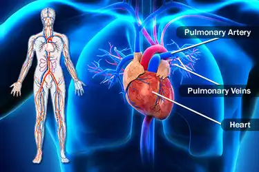
Cat Arteries & Veins Anatomym4v Watch later Share Copy link Info Shopping Tap to unmute If playback doesn't begin shortly, try restarting your device Switch cameraNew users enjoy 60% OFF 164,228,059 stock photos onlineGross anatomy There are typically four pulmonary veins, two draining each lung right superior drains the right upper and middle lobes right inferior drains the right lower lobe left superior drains the left upper lobe left inferior drains the left lower lobe The pulmonary veins course in the intersegmental septa and as such do not run with the bronchi like the pulmonary arteries do
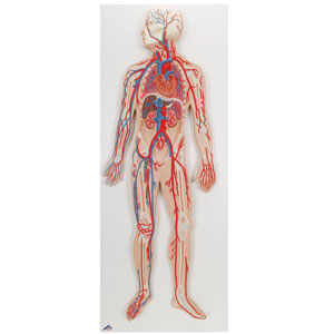
Arteries Veins Anatomy Model Ucf Libraries
Artery and vein anatomy quiz
Artery and vein anatomy quiz- There are three major types of vessels;Gross anatomy Origin and course Mesenteric venous arcades, which accompany the arteries, unite to form the jejunal and ileal veins in the small bowel mesentery and are joined by the tributaries listed below Often the superior mesenteric vein is considered the common trunk after all the chief tributaries have joined


Tunica Tunica Artery Vein Capillary Ppt Download
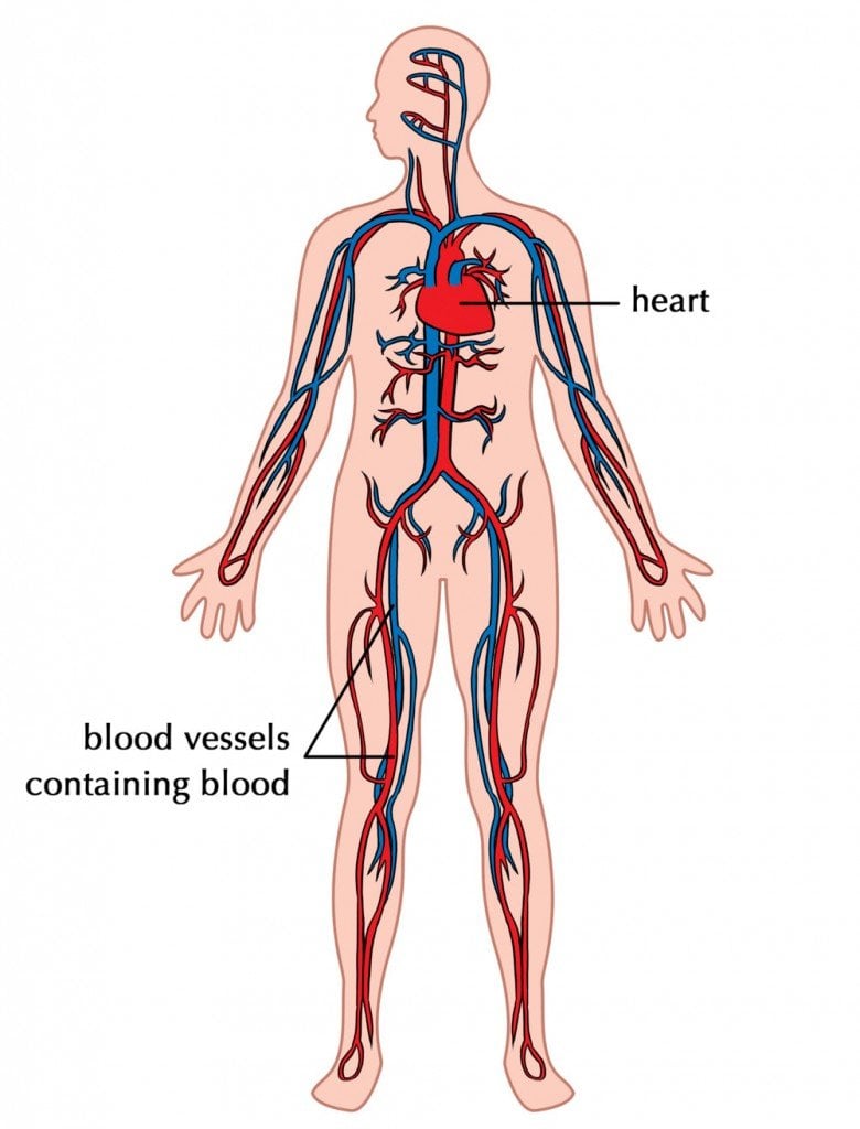
Arteries'and'Veins' ComparaveStructureof vein,'the'kidneys,'gonads'and'dorsal'por0ons'of'the' musculature''' '' '9In'fish,'the'system'is'modified'to'form'arenal'portal'system'' Blood'from'the'tail'and'posterior'trunk'flows'into'veins'called' renal'portal'veins'through'capillaries'along'the'Veins and arteries diffen science biology anatomy there are two types of blood vessels in the circulatory system of the body Artery and vein anatomy Arteries are blood vessels responsible for carrying oxygen rich blood away from the heart to the body Anatomy The median cubital vein is a part of the circulatory system Arteries, veins, and capillaries work together to carry blood, oxygen, nutrients, and waste products throughout the body Arteries carry oxygenrich blood to tissues, while veins carry blood that is depleted of oxygen and nutrients back to the heart and lungs to be replenished with more
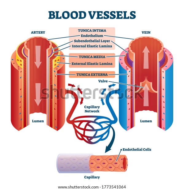
Exceptions are the pulmonary and umbilical veins, both of which carry oxygenated blood to the heartIn contrast to veins, arteries carry blood away from the heart Veins are less muscular than arteries and are often closer to the skin RENAL VASCULAR ANATOMY • The renal pedicle classically consists of a single artery and a single vein that enter the kidney via the renal hilum • The renal arteries arise from the aorta at the level of the intervertebral disk between the L1 and L2 vertebrae where the longer right renal artery passes posterior to the inferior vena cava (IVC) • Renal arteries give branches Artery, Vein, And Capillary Anatomical Structure In Detail In this image, you will find endothelium, basement membrane, internal elastic lamina, smooth muscle, external lamina, tunica external, valve, lumen, the capillary in it Our LATEST youtube film is ready to run Just need a glimpse, leave your valuable advice let us know , and subscribe us!
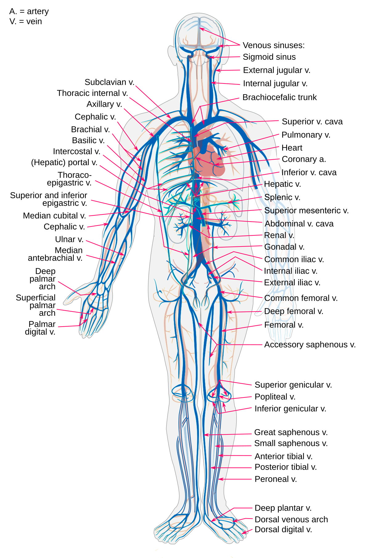
Veins of pelvis and lower limb;The internal jugular veins join with the subclavian veins to form the brachiocephalic veins,The deep veins accompany the arteries They are connected to the superficial system by perforating veins The superficial veins starts on the back of the hand as a dorsal arch •The cephalic vein begins at the radial extremity of the arch It ascends along the lateral aspect of the arm, then it pierces the deep fascia to enter the axillary vein just distal to the clavicle
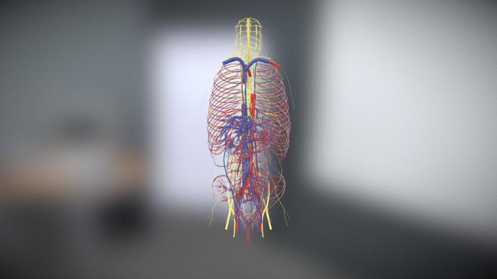


Arteries Veins Nerves And Lymphatics 3d Model By Kfiebke Kfiebke B5dbe00


1
The ventral ramus supplies the body wall and is represented in the adult by the intercostal arteries in the thoracic and lumbar arteries in the lumbar regions The dorsal aorta terminates by giving off two umbilical arteries after which it is continued by the middle sacral artery The external iliac arteries are sprouts from the umbilical arteries A new connecting trunk from the junction of the umbilical artery Posterior auricular artery Surface anatomy is represented by a line extending from the upper border of the thyroid cartilage to the neck of the mandible 4 Internal carotid artery Beginning It begins as one of the two terminal branches of the common carotid artery, opposite to the upper border of the thyroid cartilage Course It is subdivided into four parts which areDr O is building an entire video library that will allow anyone to learn Microbiology and Anatomy & Physiology for free Feel free to reach out if there ar


Anatomical Structure Of Human Body Circulatory System Arteries Veins Stock Illustration Download Image Now Istock
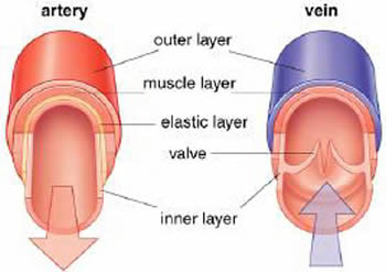


What Is The Relationship Between The Structure And Function Of Arteries Capillaries And Veins Socratic
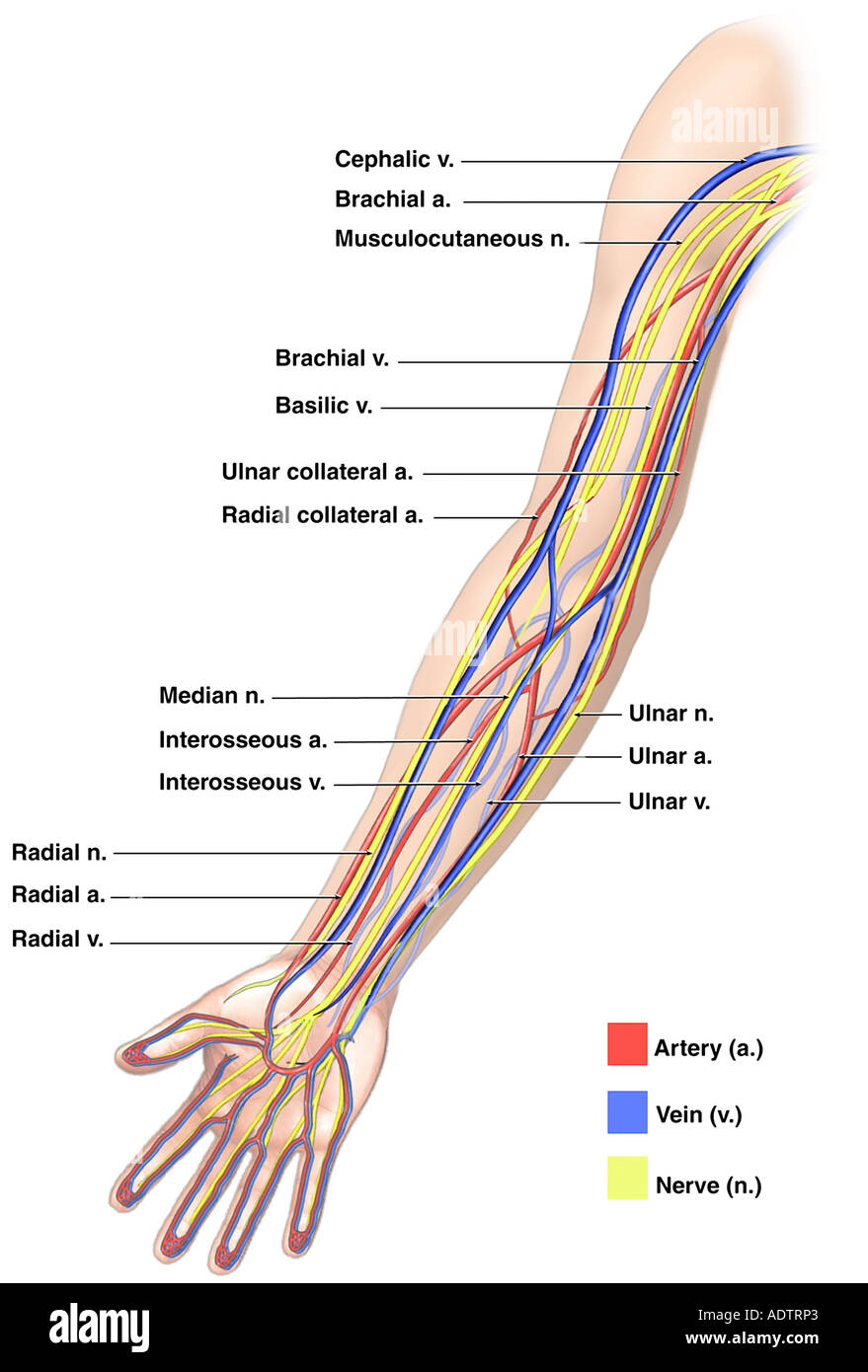
Video narrated by Professor Martin at The College of San Mateo Labels include cephalic vein, brachial artery/vein, basilic vein, musculoskeletal nerve, ulnar collateral artery note the names of the major veins and arteries involved(eg, carotid arteries and jugular veins for the head) Anatomy of the nerves, arteries and veins of the arm (upper extremity) Composition of blood arterial and venous structures head and neck There areAnatomy of arteries and veins of uterus and ovaries in rhesus monkeys Biology of reproduction, 11(2), 5219 Goldman, M P, & Fronek, A (19) Anatomy and pathophysiology of varicose veins The Journal of dermatologic surgery and oncology, 15(2), Goss, C M (1961) On anatomy of veins and arteries by Galen of Pergamos The


Biology Of The Blood Vessels Heart And Blood Vessel Disorders Msd Manual Consumer Version
:background_color(FFFFFF):format(jpeg)/images/library/11145/arteries-of-the-head-lateral-view_english.jpg)


Major Arteries Veins And Nerves Of The Body Anatomy Kenhub
They are the main path for deoxygenated blood returning from the cranium back to the heart The external jugular veins empty into the subclavian veins; Anatomy of arteries vs veins 22 right external jugular vein left common carotid artery right common carotid artery right subclavian vein left external jugular Together, veins, arteries and nerves define neurovasculature Thoracic aorta, abdominal aorta, iliac arteries veins This is an overview of the major blood vessels in the circulatory system Indicate the pathway ofArteries and veins have the same layers of tissues in their walls, but the proportions of these layers differ Lining the core of each is a thin layer of endothelium, and covering each is a sheath of connective tissue, but an artery has thick intermediate layers of elastic and muscular fiber while in the vein, these are much thinner and less developed
/the-blood-supply-of-the-pelvis-87376790-7b216233f31e4d5da3959a64e17e6a34.jpg)


External Iliac Artery Anatomy Function Significance
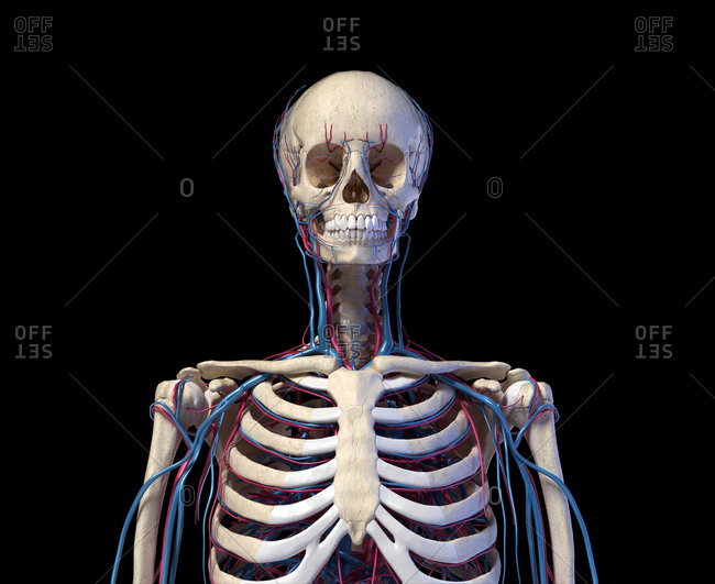


Artery Vein Stock Photos Offset
The subclavian vein (Latin vena subclavia) is a large blood vessel that arises from the axillary vein It is one of the deep veins of the neck The subclavian vein originates at the outer border of the first rib It travels within the subclavian groove, then runs laterally to the medial border of the anterior scalene The umbilical vein arises from multiple small veins within the placenta which carry oxygen and nutrient rich blood derived from the maternal blood circulation via the chorionic villi From here, it enters the umbilical cord, along with the paired umbilical arteries After emerging from the umbilical cord into the abdominal cavity of the fetus, it passes within the layers of theThe superficial veins of the lateral aspect of the foot join together to form the short saphenous vein The ones on the medial aspect of the foot join together to form the long saphenous vein In addition, at a deeper level, the arteries, which we'll be looking at next, are closely accompanied by concomitant veins, like these From here on we


Anatomy Artery Veins Arterial Vein Blood Stock Vector Royalty Free
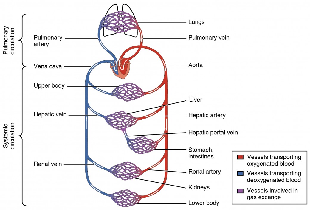


Structure And Function Of Blood Vessels Anatomy And Physiology Ii
Browse our range of artery anatomy models and charts, to learn about the structure and function of these important blood vessels Enlarged models help educate viewers about arterial health, and show the effects of diet and smoking on the body and how arteries can become clogged by cholesterol and plaque Use a functional artery model to engage viewers about pathologies affecting the veinsAnatomy_of_femoral_artery_and_vein 3/18 Anatomy Of Femoral Artery And Vein Circulation through the deep femoral artery and its branches is critical to patients with aortoiliac and infrainguinal arteriosclerosis It is, accordingly, essential that all physicians who are seriously interested in treating patients with lower extremity ischemia have a good working knowledge of this crucial artery Veins do not contain the elastic membrane lining that is found in arteries In some veins, the tunica intima layer also contains valves to keep blood flowing in a single direction Vein walls are thinner and more elastic than artery walls This allows veins to hold more blood than arteries


Tunica Tunica Artery Vein Capillary Ppt Download
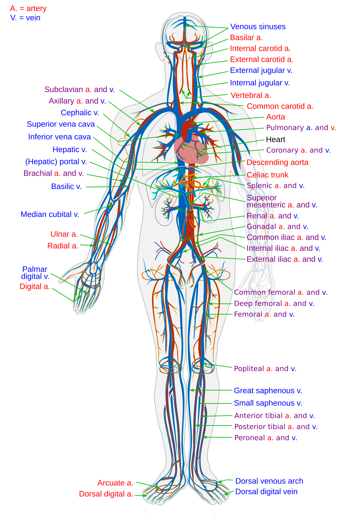


Blood Vessel Wikipedia
Arteries, veins and capillaries Arteries (with the exception of the pulmonary artery) deliver oxygenated blood to the tissues At the tissues, the oxygen and nutrient exchange is carried out by the capillaries The capillaries also return deoxygenated blood to the veins, which bring it back to the heart (with the exception of the Tunica Intima the inner layer of arteries and veins In arteries, this layer is composed of an elastic membrane lining and smooth endothelium (a special type of epithelial tissue) that is covered by elastic tissues The artery wall expands and contracts due to pressure exerted by blood as it is pumped by the heart through the arteries Arterial expansion andAnatomy of the Nerves, Arteries and Veins of the Arm (Upper Extremity) Labels include cephalic vein, brachial artery/vein, basilic vein, musculoskeletal nerve, ulnar collateral artery, radial collateral artery, ulnar nerve/artery/vein, interosseous artery/vein, median nerve and radial nerve/artery/vein Save up to 30% with our image packs Prepay for multiple images and
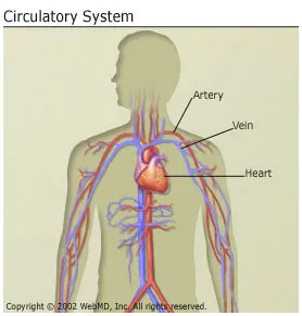


Anatomy And Circulation Of The Heart
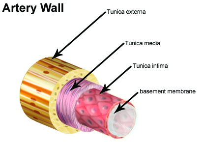


Seer Training Classification Structure Of Blood Vessels
Arteries and veins of the orbit (or eye) are generally thought of as the central retinal artery and retinal vein, in addition to the ophthalmic artery and vein However, there are a number of additional ancillary arteries and veins that help support proper structure and functioning of Veins are the large return vessels of the body and act as the blood return counterparts of arteries Because the arteries, arterioles, and capillaries absorb most of the force of the heart's contractions, veins and venules are subjected to very low blood pressures This lack of pressure allows the walls of veins to be much thinner, less elastic, and less muscular than the walls of arteriesKizzlethadon artery/vein anatomy STUDY PLAY vascular supply the main blood supply to the head and neck is from the _______ and _______ arteries(the origins of these arteries differs for the right and left sides) 1 subclavian 2 common carotid


Venule Anatomy Britannica
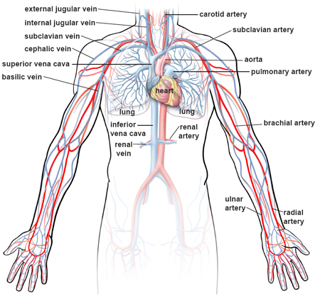


Illustrations Of The Blood Vessels
Middle hepatic vein This vein runs at the middle portal fissure, dividing the liver into right and left lobes It runs just behind the IVC It runs just behind the IVC Left hepatic vein This vein is found in the left portal fissure, splitting up the left lobe of Arteries are a type of blood vessel They work to carry blood away from the heart In contrast, veins carry blood back to the heart Because arteriesBlood is transported in arteries, veins and capillaries Blood is pumped from the heart in the arteries It is returned to the heart in the veins The capillaries connect the two types of blood


Healthy Artery And Vein Anatomy Layers Of Arteries And Veins Medical Illustration Royalty Free Cliparts Vectors And Stock Illustration Image


Veins Of The Body Part 1 Anatomy Tutorial Youtube
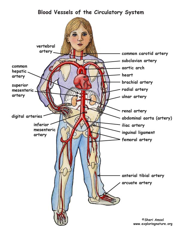
Arteries thoracic aorta, abdominal aorta, iliac arteries Veins superior vena cava, azygos, hemiazygos, iliac veins, inferior vena cava Nerves medial pectoral, lateral pectoral, intercostal, subcostal, phrenic, vagus, pelvic splanchnic nerves, lumbar plexus (L1L4) Upper extremity Arteries axillary, brachial, ulnar and radial arteries The middle layer of the walls of arteries and veins is called the tunica media It's made of smooth muscle and elastic fibers This layer is thicker in arteries and thinner in veins InnerThe artery and vein model shows a mediumsized muscular artery with two adjacent veins from the antebrachial area with adjoining fat tissue and muscle enlarged 14 times The MICROanatomy™ circulatory system model illustrates the reciprocal anatomical relationship of artery and vein and the basic functional techniques of the venous valves ("valve function" and "muscle pump") The left vein and the middle artery


Free Vector Arteries And Veins Of The Leg


Coronary Arteries And Cardiac Veins Preview Human Anatomy Kenhub Youtube
The outermost layer of an artery (or vein) is known as the tunica externa, also known as tunica adventitia, and is composed of collagen fibers and elastic tissue with the largest arteries containing vasa vasorum (small blood vessels that supply large blood vessels)Explore Jeanine Inciriaga's board "Arteries and veins" on See more ideas about arteries and veins, arteries, anatomy and physiologyYour Anatomy Artery Vein stock images are ready Download all free or royaltyfree photos and vectors Use them in commercial designs under lifetime, perpetual
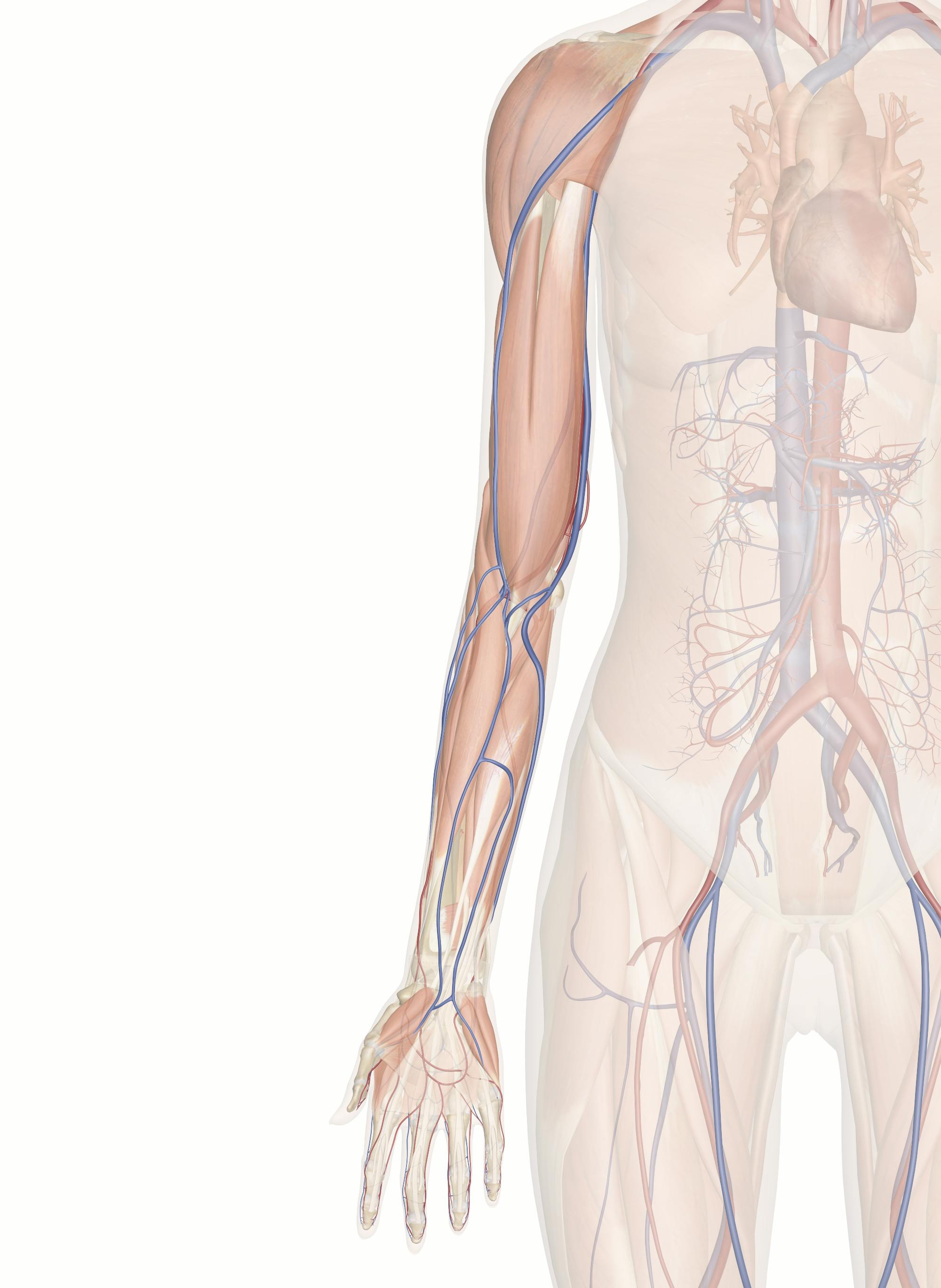


Cardiovascular System Of The Arm And Hand
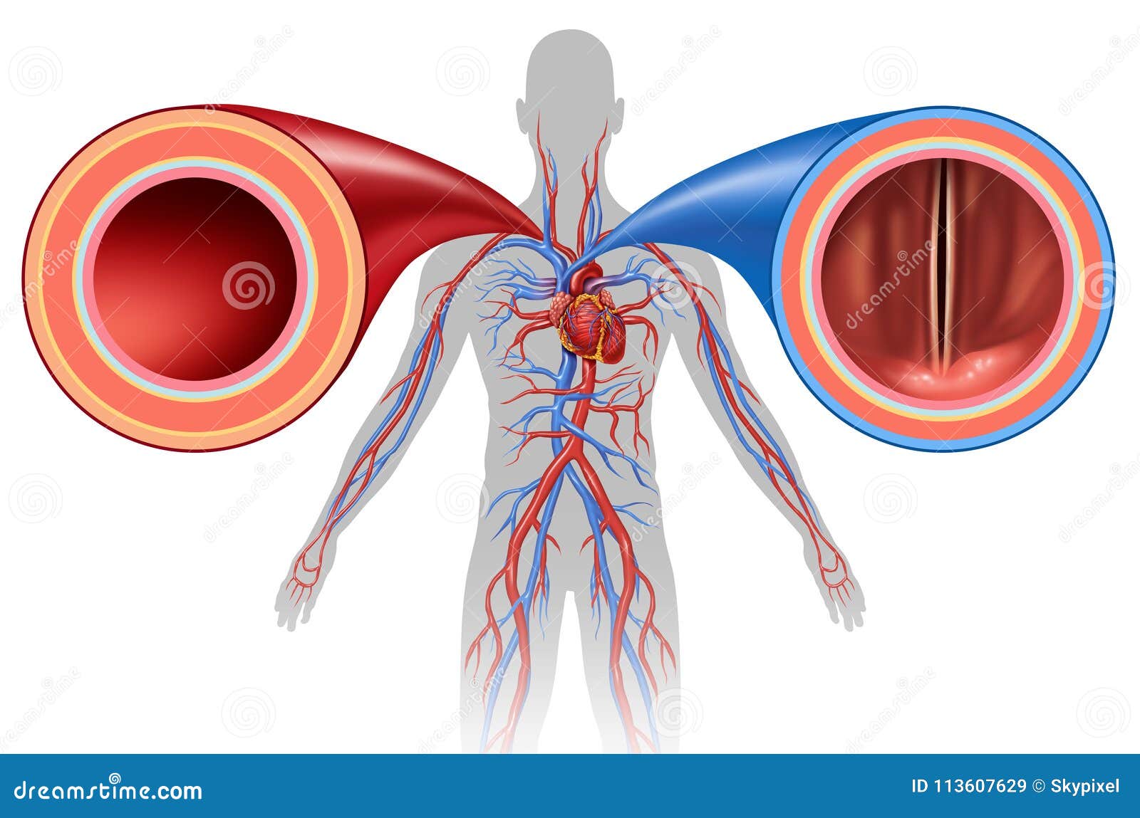


Artery And Vein Human Circulation Stock Illustration Illustration Of Circulatory Anatomy
Veins And Arteries Of The Leg diagram and chart Human body anatomy diagrams and charts with labels This diagram depicts Veins And Arteries Of The LegHuman anatomy diagrams show internal organs, cells, systems, conditions, symptoms As the internal jugular vein runs down the lateral neck, it drains the branches of the facial, retromandibular, and the lingual veins The course of the internal jugular vein is directed caudally in the carotid sheath, accompanied by the vagus nerve posteriorly and the common carotid artery anteromedially It lies just lateral and anterior to the internal and common carotid arteries At the junction of the neck and thorax, the internal jugular vein The vessels supplying the lungs include the pulmonary arteries, pulmonary veins, and bronchial arteries The segmental and sub segmental pulmonary arteries parallel the bronchi and are named according to the bronchopulmonary segments they supply There are however considerable anatomic variations, particularly in the upper lobes with variations in number or presence of accessory arteries from adjacent segments The subsegmental pulmonary vein


Body Arteries Veins 3d Turbosquid
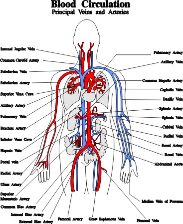


Blood Vessels Arteries Capillaries Veins Vena Cava Central Veins Lhsc
Download 14,970 Anatomy Artery Vein Stock Illustrations, Vectors & Clipart for FREE or amazingly low rates!Femoral Blood Vessels (Artery and Vein), Anatomy, Pictures Posted by Dr Chris The femoral blood vessels are important conduits for blood traveling between the heart and lower limb The femoral artery carries blood to the lower limb while the femoral vein carries blood back to the heart These structures are common sites for conditions that cause narrowing or blockage of the3B MICROanatomy™ Artery & Vein Model, 14 times Enlarged 3B Smart Anatomy Human Heart Models 3B MICROanatomy Artery & Vein Model, 14 times Enlarged , includes 3B Smart Anatomy, the 3D human anatomy course for virtual learning 3B Scientific offers highquality handson student and patient education anatomical models


A Arterial Anatomy At The Fingertip B Dorsal Venous Anatomy At The Download Scientific Diagram
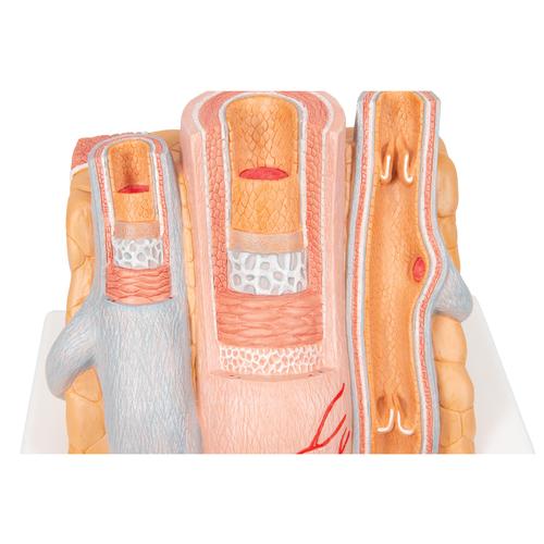


Anatomical Artery Vein Model Anatomy Of The Artery Vein Circulatory System Model Microanatomy Model
Veins are blood vessels that carry blood towards the heartMost veins carry deoxygenated blood from the tissues back to the heart;Anatomy of arteries and veins of submandibular glands Chin Med J (Engl) 07 Jul 5;1(13)1179 Authors Li Li 1 the venae comitantes of facial artery, the vein close to the Whaston's duct (the hilum vein), and seldom drained to external jugular vein and other veins Conclusions The anatomy of SMG is a complicated structure Determining the main blood
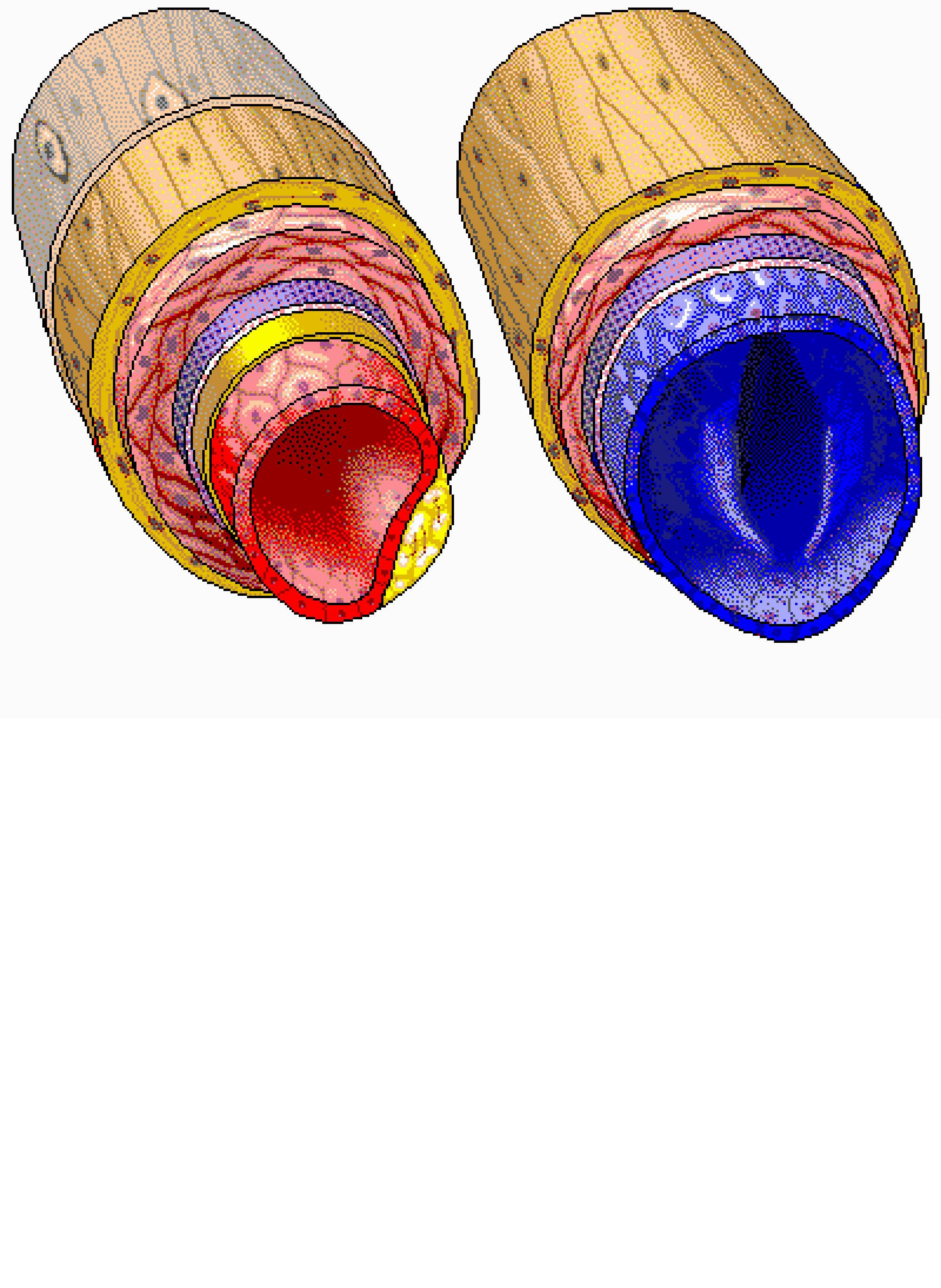


Cross Section Of Artery And Vein Interactive Anatomy Guide


Circulatory System Human Anatomy Diagram On Female Body With Arteries And Veins Stock Illustration Download Image Now Istock
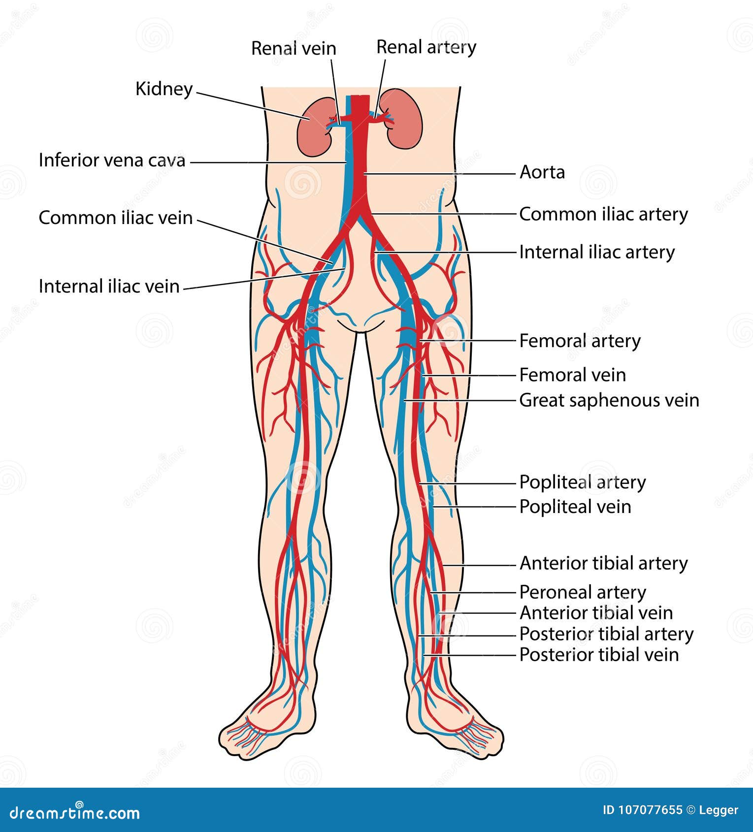


Blood Vessels Of The Lower Body Stock Vector Illustration Of Arteries Vein
:background_color(FFFFFF):format(jpeg)/images/library/12609/intro.png)


Intercostal Arteries And Blood Supply Of Thoracic Wall Kenhub


Anatomy Of The Nerves Arteries And Veins Of The Arm Upper Extremity Labels Include Cephalic Vein Brachial Ar Nerve Anatomy Arteries And Veins Median Nerve


Capillary Pulmonary Artery Vein Function Heart Heart Lung Anatomy Png Pngwing
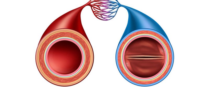


Difference Between Arteries And Veins Pva Doctors Of Arteries Veins
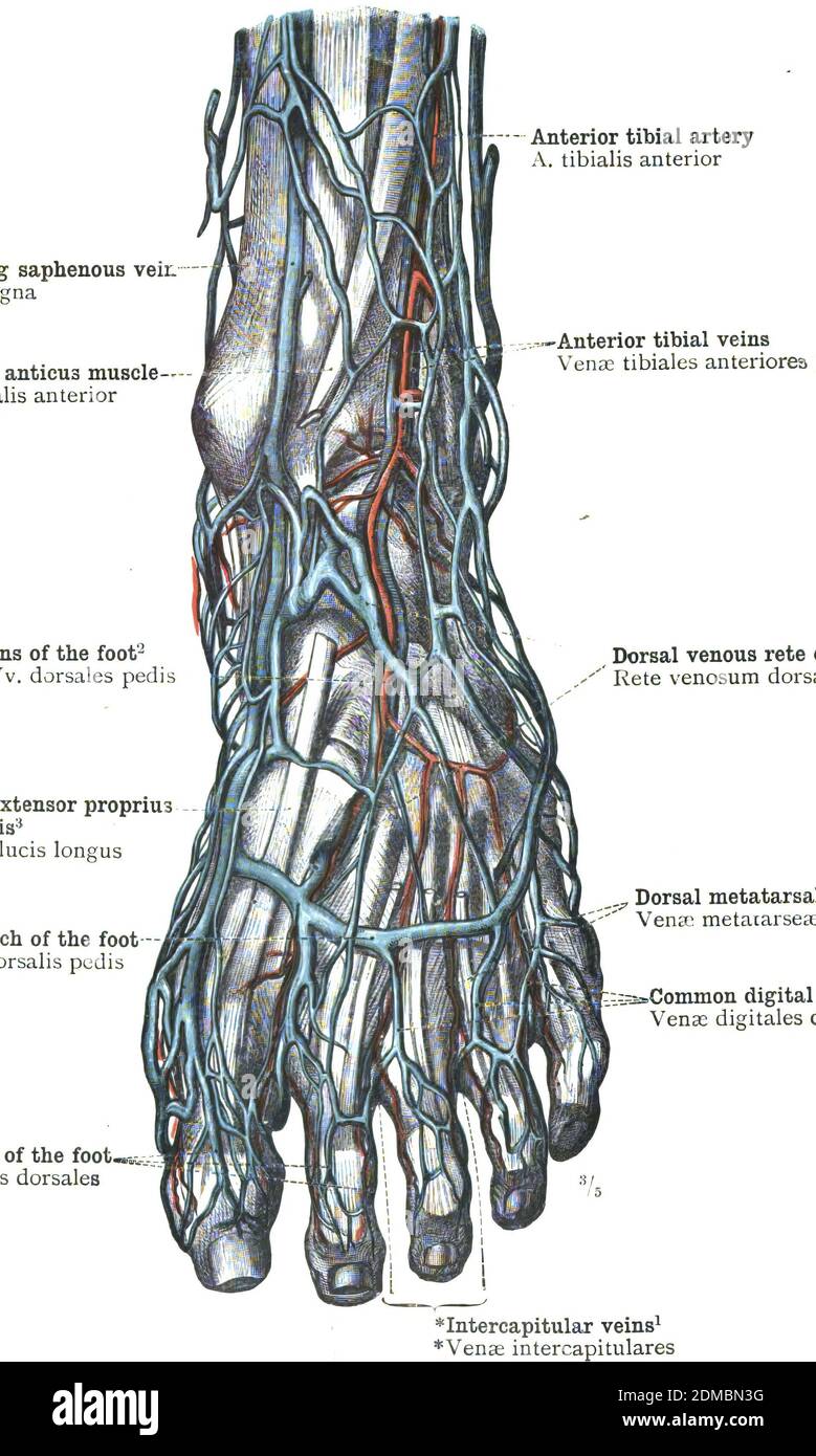


Veins Arteries Foot High Resolution Stock Photography And Images Alamy
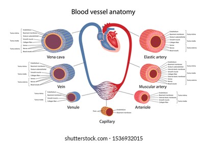


Artery Vein Images Stock Photos Vectors Shutterstock


1


Arteries Veins And Nerves Of The Upper Arm And Shoulder Preview Human Anatomy Kenhub Youtube
:background_color(FFFFFF):format(jpeg)/images/library/13977/Coronary_vessels_cardiac_veins.png)


Coronary Arteries And Cardiac Veins Anatomy And Branches Kenhub
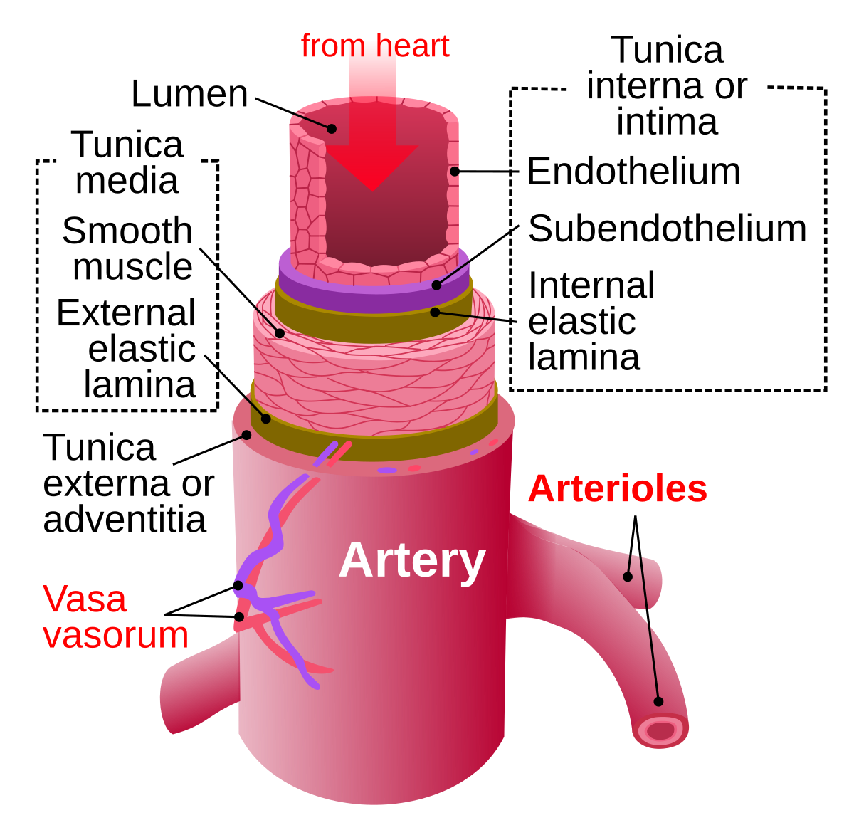


Artery Wikipedia
:background_color(FFFFFF):format(jpeg)/images/article/en/neurovasculature-of-head-neck/DUdNRgQUmaTFMGS6j2Y79Q_Neurovasculature_of_head___neck.png)


Nerves And Arteries Of Head And Neck Anatomy Branches Kenhub
:background_color(FFFFFF):format(jpeg)/images/library/11144/pasted_image_0__1_.png)


Major Arteries Veins And Nerves Of The Body Anatomy Kenhub


How To Do Venous Blood Sampling Critical Care Medicine Msd Manual Professional Edition


Brachial Veins Wikipedia
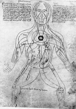


History Of The Arteries And Veins
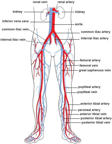


Illustrations Of The Blood Vessels
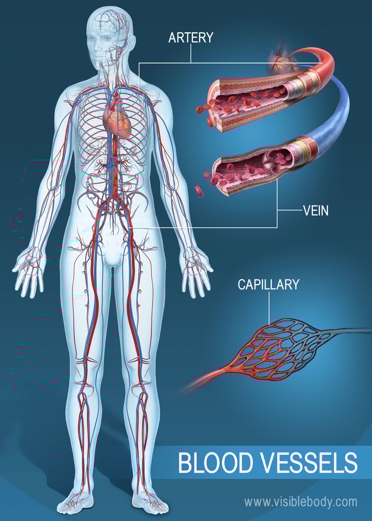


Blood Vessels Circulatory Anatomy


What Is The Difference Between Arteries Veins Nerves Youtube


Arteries Veins Anatomy Model Ucf Libraries


1
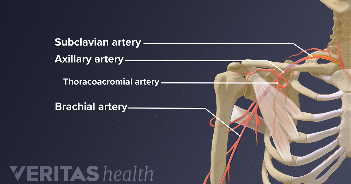


Blood And Nerve Supply Of The Shoulder
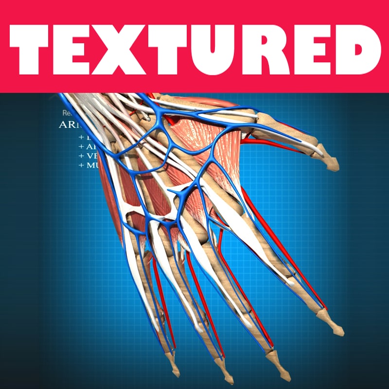


Anatomy Arteries Veins 3d Model


Healthandfitnessmagazine Leg Vein Anatomy Medical Anatomy Arteries And Veins


Blood Vessel Structure And Function Boundless Anatomy And Physiology


Artery Vein And Capillary Anatomical Structure In Detail
/GettyImages-87302280-83604c7a3ca84315a84304a002377404.jpg)


Femoral Vein Anatomy Function And Significance
:background_color(FFFFFF):format(jpeg)/images/library/11146/ventral-trunk-nerves-vessels_english.jpg)


Major Arteries Veins And Nerves Of The Body Anatomy Kenhub


Blood Vessels Artery Vein Internal Structure Stock Vector Royalty Free
/arterial_system-59a5bdab68e1a200136f1b53.jpg)


Artery Structure Function And Disease


Arteries Veins Atlas Of Anatomy


Human Being Anatomy Blood Circulation Principal Veins And Arteries Image Visual Dictionary


Visual Guide To Vein And Artery Problems


Arteries Veins Atlas Of Anatomy


Arteries Or Veins What S The Difference


01x Arteries And Veins Anatomy Exhibits


Mesenteric Artery Anatomy Britannica


Fig Forearm And Hand Arterial And Venous Anatomy With The Autogenous Download Scientific Diagram


Arteries Veins And Capillaries Access Revision
:background_color(FFFFFF):format(jpeg)/images/library/11147/upper-arm-nerves-vessels_english.jpg)


Major Arteries Veins And Nerves Of The Body Anatomy Kenhub


Anatomy Of Thoracic Outlet The Subclavian Artery Vein And Brachial Download Scientific Diagram


Anatomical Structure Human Bodies Blood Vessels With Arteries Veins Stock Vector Illustration Of Bloodstream Body


Arteries Veins Capillaries Diagram High Res Stock Images Shutterstock


Anatomy Of The Nerves Arteries And Veins Of The Arm Upper Extremity Stock Photo Alamy


Blood Vessels Arteries Veins And Capillaries


What Are Arteries Veins And Capillaries Science Abc


Vein Wikipedia
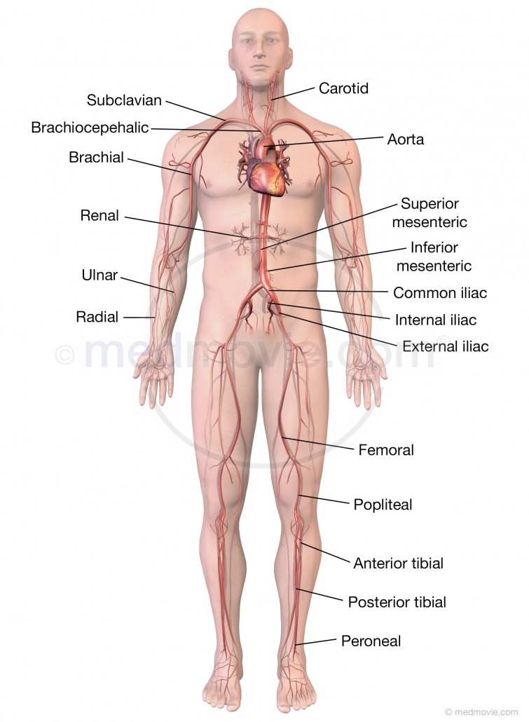


Major Arteries Of The Body Medmovie Com


Pin On Health Issues


Apii Notes Home Page


Interior View Of Human Chest Heart Lungs Arteries Veins Anatomy Stock Photo Download Image Now Istock
/vascular-system-veins-56c87fa03df78cfb378b3e7c.jpg)


What Is A Vein Definition Types And Illustration


Dysautonomia Pots Awareness Dysautonomia Pots Awareness Support Circulatory System Arteries Arteries And Veins


Rectal Artery An Overview Sciencedirect Topics


Arteries Boundless Anatomy And Physiology


Human Being Anatomy Blood Circulation Principal Veins And Arteries Image Visual Dictionary Online


Vein Png Images Pngegg
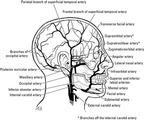


Veins Arteries And Lymphatics Of The Face Dummies


Blood Vessels Circulatory Anatomy
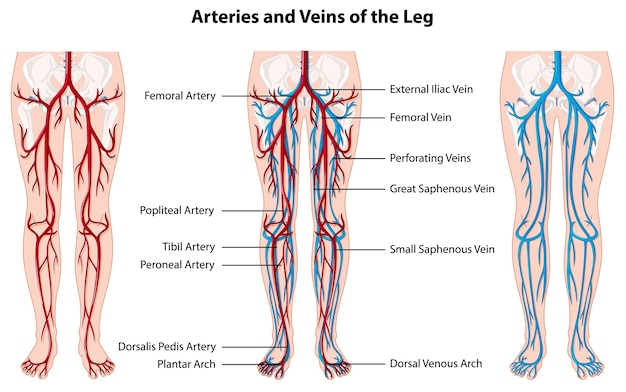


Free Vector Arteries And Veins Of The Leg


Human Circulatory System Of Arteries And Veins Poster Zazzle Com In 21 Human Circulatory System Arteries And Veins Circulatory System
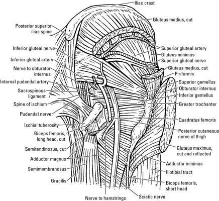


Arteries Veins And Lymph In The Hip And Thigh Dummies


What Is The Difference Between An Artery And A Vein
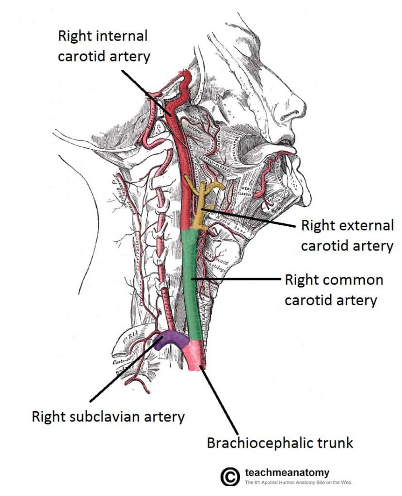


Major Arteries Of The Head And Neck Carotid Teachmeanatomy


Ultrastructure Of Blood Vessels Arteries Veins Teachmeanatomy


Anatomy Of Arteries Veins And Capillaries Ppt Video Online Download


Theory Of Compression Altimed Compression Hosieryaltimed Compression Hosiery


Artery Vein Stock Photos Offset


1


Renal Vein Anatomy Britannica
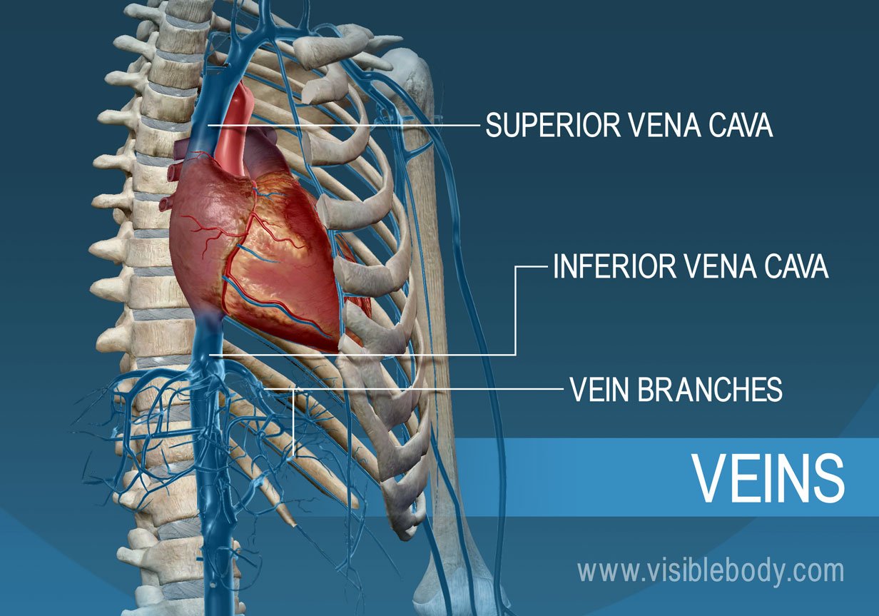


Blood Vessels Circulatory Anatomy


Coronary Circulation Wikipedia


Pulmonary Artery Anatomy Britannica


Your Heart Blood Vessels


How To Distinguish Between Artery Vein And Nerves In A Cadaver During Dissection Quora


Blood Vessels Biology For Majors Ii


0 件のコメント:
コメントを投稿